Nervous system (of man). -
I. General considerations
It is a general property of protoplasm to be excitable, that is, to perceive stimuli of various kinds and to react to them with appropriate responses. In unicellular animals, the protozoa, this function is performed by the single cell that constitutes the individual, which, however, may exhibit particular differentiations in this regard.
II. DEVELOPMENT OF THE NERVOUS SYSTEM IN VERTEBRATES AND MAN
The nervous system originates from an extremely early embryonic anlage that becomes apparent at the time of gastrulation: the neural plate. As the plate folds, the neural tube is formed, which initially is open anteriorly (anterior neuropore) and posteriorly communicates with the primitive gut (neuroenteric canal).Of great importance for the subsequent morphogenesis of the neural tube is the condition of the notochord, which forms beneath II. This supporting organ does not extend to its cephalic end but terminates earlier, so that the neural tube is divided into a precordal part and a chordal part. The precordal part, lacking the chordal obstacle, dilates into vesicles, as does, though much more moderately, the most cephalic part of the chordal portion; the remainder of the chordal part does not dilate and will give rise to the spinal cord, while the vesicles will give rise to the brain. After a two-vesicle stage and an intermediate three-vesicle stage, the five-vesicle stage is reached (the telencephalon, diencephalon, mesencephalon, metencephalon, and myelencephalon).
From this stage, characteristic in all vertebrates, the definitive differentiation proceeds through the development of two sacciform portions on the sides of the telencephalic vesicle, which in their maximal development will represent the cerebral hemispheres, and through the unequal thickening of the walls of the various vesicles, which also leads to a different width of the internal ventricular cavities. The spinal part of the neural tube, through the thickening of its lateral walls, is transformed into the spinal cord, and its internal cavity is reduced to a slender canal, the ependymal canal. The development of the cerebral walls varies in the different classes of vertebrates in that it becomes more complex as one ascends the zoological scale, reaching the most conspicuous structural differentiations in man. The thickening of the walls of the neural axis is brought about by the proliferation of the ependymal germinal layer, which produces two types of undifferentiated cells, the "neuroblasts" and the "spongioblasts," which migrate from this site to the exterior, where they respectively differentiate into nerve cells and neuroglial cells. During their migration, guided by stimuli of various kinds, the neuroblasts gather into nuclei and nerve centers.
III. MORPHOLOGY OF THE NERVOUS SYSTEM IN VERTEBRATES AND MAN
In the spinal cord the nerve cells form cellular columns (grey matter) along the length of the cord, which in cross-section assume the shape of an H or a butterfly with open wings; two dorsal horns and two ventral horns can thus be identified.According to the research of Johnston, four functional areas can be distinguished in the spinal cord: in the most apical part of the dorsal horns lies the somato-sensory area, where somatic sensations arrive; in the most apical part of the ventral horns lies the viscero-motor area, from which motor impulses depart for the skeletal musculature; between these two areas, in an intermediate position, more dorsally, there is the viscero-sensory area, where visceral sensations arrive, and the somato-motor area, from which motor impulses depart for the visceral musculature. The efferent motor fibres exit from the ventral part and gather into bundles of fibres that form the ventral roots of the spinal nerves; the afferent sensory fibres reach the spinal cord from the dorsal part (dorsal roots) and along their course the spinal ganglia are situated. The two roots unite into mixed spinal nerves. The sensory fibres that enter the spinal cord partly connect with cells of the dorsal horns and partly proceed cranially, reaching cell groups located in the medulla oblongata (nuclei of the fasciculus gracilis and the fasciculus cuneatus). From the cells of the spinal sensory nuclei fibres depart that reach various zones of the medulla oblongata (or bulb), the midbrain, the cerebellum, and the thalami of the diencephalon. The nervous element of the spinal ganglia, a typical bipolar element in the embryo (later transforming into a T-shaped cell), thus provided with two prolongations, on one side connects with the spinal cord as described above, and on the other establishes relations with the periphery through various peripheral devices (expansions, diffuse networks) and sensory corpuscles (of Meissner, Pacini, Golgi-Mazzoni, etc.).
Anterior to the spinal cord lies the brain with its various subdivisions. The encephalic portion that connects directly with the spinal cord is the medulla oblongata or bulb, which still closely resembles the spinal cord in morphology. The medulla oblongata presents thickened lateral walls and base, while the roof remains thin and forms the choroid tela of the fourth ventricle, represented by a wide dilation of the ependymal cavity deriving from the myelencephalic vesicle.
In addition to the named centres for the connection of the long ascending pathways from the spinal cord, the bulb contains motor and sensory centres of cranial nerves and centres for motor coordination (tegmental centres, inferior olives). There are motor nuclei of somatic nerves such as the hypoglossal (XII pair) and the abducens nerve of the eye (VI pair), and the nuclei of the visceral nerves that innervate musculature of branchial origin, the salivary glands, and the smooth musculature of the body's viscera, such as the glossopharyngeal (IX pair), the vagus (X pair), and the accessory (XI pair), and the somato-sensory nuclei of the trigeminal (V pair), the cochlear and vestibular nerves, which constitute the acoustic complex (VIII pair), and the viscero-sensory nuclei of the VII, IX, and X pairs that form the so-called “solitary fasciculus.” In the white matter of the bulb the passage of the long pathways that go from the spinal cord to the higher centres and of those descending from these to the spinal cord is found. Anterior to the medulla oblongata are the differentiations of the metencephalic vesicle, which, with the development of the dorsal part, give rise to the “cerebellum.” In this structure centres differentiate that connect on one side with general proprioceptive sensitivity (joint, tendon, muscular sensitivity) and special sensitivity (semicircular canals) and on the other with all the motor impulses of the cortical, pyramidal, and extrapyramidal pathways, becoming the central organ for the coordination and synergism of movement and for the integration of all motor and equilibrium activities. It acts autonomously, outside of voluntary control, though it is connected with the cerebral cortex via the centres of the pons of Varolius. The cerebellar response occurs through the cerebellar olives or dentate nuclei and the reflex motor centres of the tectum and the red nucleus of the midbrain.
Anterior to the midbrain, the mesencephalon splits in all its walls so that the ventricular cavity is reduced to a slender canal known as the “aqueduct of Sylvius.” In its basal part other motor centres of the eyes are found (the oculomotor, III pair, and the trochlear, IV pair) and some centres of the extrapyramidal motor pathway (of which more will be said shortly), such as the red nucleus and the substantia nigra. Moreover, the long ascending and descending pathways that do not have a station in this sector pass through the midbrain. The dorsal part is constituted by the quadrigeminal plate, formed by a pair of anterior colliculi, reflex centres for vision, and a pair of posterior colliculi, reflex centres for hearing. The diencephalic vesicle, anterior to the midbrain, with development becomes differentiated into the thalami, thickenings of the lateral walls, the hypothalamus, a thickening of the base, and the epithalamus, a thickening of the roof. The cavity is reduced to a wide vertical fissure that, together with the unpaired cavity of the subsequent telencephalic vesicle, forms the third ventricle.
The thalami are for the most part sensory centres where the long pathways coming from the spinal cord and other posterior sectors (bulb, midbrain) arrive; a ventral portion (subthalamus) is motor. In the sensory part of the thalami the following are distinguished: an optic zone (pulvinar) that connects with a differentiation of the thalamus itself, the lateral geniculate body; an acoustic zone, connected with the medial geniculate body; a zone for cutaneous sensitivity, coming from the long exteroceptive pathways of the spinal cord and the bulb. In the thalamus there are also association centres that, in lower vertebrates lacking a cerebral cortex, represent the highest centres where the most complex nervous activities are elaborated, though of a primitive nature, such as instincts, in which no expression of conscious will is manifested.
The most anterior vesicle, the telencephalon, is the one that undergoes the greatest differential complication and is the one that presents the widest degrees of development in the series of vertebrates. This vesicle, initially unpaired and median like the others, evaginates into two lateral vesicles so that two lateral ventricles (I and II) are formed. In lower vertebrates (fish, amphibians, and lower reptiles) these hemispheres are exclusively related to olfactory sensitivity (paleopallium and archipallium); at the base, thickenings develop, known as basal ganglia or striate bodies, which represent the most cephalic motor station and which, even in higher forms, represent a centre of the extrapyramidal pathway.
In the study of the complexity of brain structures, the question of the crossing of fibres must first be considered. Indeed, the great majority of nerve fibres that have a peripheral relationship with one side are connected to centres on the opposite side; thus along the median sagittal zone of the neuraxis, decussations, chiasmata or commissures are observed, which sometimes constitute very conspicuous anatomical entities. In the telencephalon, three main commissures are noted that contribute greatly to its morphology: the anterior commissure
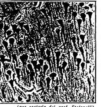
NERVOSO, SISTEMA (DELL'UOMO) - Cellule piramidali della corteccia cerebrale.
The inferior commissure and the hippocampal commissure are exclusively composed of olfactory pathways (and are therefore the most primitive), while the corpus callosum carries the decussation pathways of the neocortex (and is thus the most evolved commissure). The corpus callosum reaches its greatest development in man, since in him the neocortex also attains its maximum development. With the development of the corpus callosum, the olfactory cortex is pushed far back from its initial morphological position, so that the fibers forming the hippocampal commissure must travel a long course along the roof of the third ventricle, thereby constituting that anatomical structure known as the "three-pillared vault" or "fornix."
The development of the cerebral cortex is primarily in surface area, and in man it presents a very large number of folds (convolutions). In the human species, the greatest complexity is found, far surpassing even the most highly evolved mammals, such as the anthropoid apes (v. CERVELLO). The deepest sulci divide the cerebral cortex into five lobes: occipital, temporal, frontal, parietal, and the insula, the latter lying deep and being operculated by the temporal lobe, which has grown over II. The sulci delimit the gyri.
IV. PHYSIOLOGY
A fundamental subdivision of the nervous system is that into two subsystems, which are distinct both morphologically and physiologically, though in intimate anatomical-functional connection: a “somatic” nervous system, which participates in the activities of the life of relation, and a “visceral” or “neurovegetative” nervous system (see below, § V), which participates in the physiological activities of the viscera. While the somatic nervous system operates through involuntary and voluntary mechanisms, the visceral nervous system acts exclusively in an autonomous or involuntary manner.The somatic nervous system is represented by its peripheral part (peripheral sensory organs, motor end-plates, peripheral nerves, spinal ganglia) and by a central part (the somatic portions of the spinal cord and brain); the visceral nervous system is formed by peripheral sensory organs, nerves, visceral and spinal ganglia, and by the central visceral parts of the spinal cord and brain (the presence of visceral centers in the telencephalic portion alone is still debated). The viscera have a double visceral innervation, sympathetic and parasympathetic, with antagonistic action. The adequate function for the physiological moment is maintained by the equilibrium of these two activities.
V. RECEPTORS AND EFFECTORS
Nervous activity is manifested through the excitability of receptors and the activity of effectors. Receptors consist either of sensory cells located in specific peripheral epithelia and the sensory ganglion neuron that transmits excitation to the central nervous system, or of ganglionic cells themselves whose peripheral prolongation is complicated by microscopic peripheral devices (corpuscles, networks, plexuses, arborizations, etc.) situated in epithelia or connective tissues. Effectors consist of the motor nerve cell (spinal, bulbar, mesencephalic, or ganglionic), its peripheral prolongation, which becomes complex in terminal apparatuses adhering to striated muscle fibers (motor end plates) or smooth muscle fibers (with various arborizations).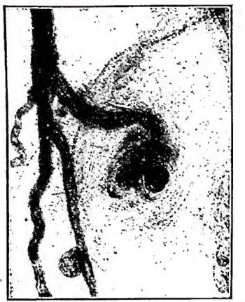
Receptors can be somatic or visceral. Somatic receptors, according to Sherrington, are subdivided into exteroceptors and proprioceptors: the former are responsible for directly perceiving stimuli from the external world (tactile, thermal, visual, acoustic, olfactory stimuli), while the latter do so indirectly insofar as the site of stimulation is in the joints, tendons, and muscles, in relation to the stance on the limbs and muscular efforts, activities that are indirectly related to the environment; to these must be added the excitability of the receptors of the semicircular canals, which, together with the aforementioned muscular receptors, give the sensation of the position of the body relative to the environment, which represents the perception necessary for the function of equilibrium. Visceral receptors are called visceroreceptors and are represented by the neurons of the visceral nervous system and their peripheral expansions (recti, plexuses, and corpuscles). The effectors, consisting of the various motor units, are also somatic, represented by the somatic motor neuron and the striated muscle fibers connected to it by the terminal motor end plates, and visceral, represented by the ganglionic visceral motor neuron and its expansions on the smooth or striated (heart) muscles of the viscera to which it is connected.
Of particular physiological importance are the contact zones between the peripheral sensory cell (where it exists) and the sensory fiber and that between the motor end plate and the muscle fiber, called "synapses," and the relationships between neuron and neuron in the constitution of chains (interneuronal synapses). The synapse allows the transmission of excitation while preserving the integrity of the anatomical unit (neuron theory), permits through its multiplicity the "spatial summation" of excitation, explains the latency period in reflex conduction, and explains the polarization of conduction in a chain of neurons. The mechanism by which excitation crosses the synapse has been the subject of much research, and today it is considered from a physical point of view (production of action potentials, depolarizing the membrane of the neuron in which the excitation is transmitted, etc.) and from a chemical point of view for the production of "chemical mediators" (acetylcholine, inactivated after the action of cholinesterase).
VI. REFLEXES
It has already been noted in the study of the evolution of the nervous system how the simplest nervous mechanism is expressed through the intervention of only two neurons: the sensory neuron and the motor neuron. This is called a simple reflex arc. The spinal cord is the seat of these reflexes by means of an arc comprising the sensory neuron, whose cell body is in the spinal ganglion, and the motor neuron whose cell body is in the anterior horns, peripherally connected with the fibers of a muscle. This spinal reflex arc, like all reflexes, is involuntary. In the spinal cord, extensor reflexes are manifested (such as the well-known knee-jerk reflex, elicited by tapping below the patella) and flexor reflexes (such as those due to painful stimuli at the extremities). These two associated reflexes are responsible for maintaining the animal on its limbs in opposition to gravity. When a muscle contracts, the antagonistic apparatus is released according to a fundamental principle of reciprocal innervation and inhibition. More complex spinal reflex mechanisms are the intersegmental ones, by which the reflex movement of a pair of limbs involves an "integration" with movements of other muscles of the trunk or of the other pair of limbs, and even with effects on the visceral sphere (genital organs). This is clearly demonstrated in spinal animals, i.e., those with the spinal cord severed and separated from the brain.The physiological value of the higher brain centers on spinal activities can be studied by shifting the plane of resection more and more cephalad, or by removing the brain with resections at increasingly higher levels.
If a resection is performed before the pons of Varolius, the characteristic decerebrate rigidity appears, which is caused by a continuous contraction of the muscles, predominantly extensors. This occurs due to the continuous excitation of the vestibular centers, which remain connected to the spinal cord and are not dominated by the higher centers, particularly the extrapyramidal system. Postural, static movements are expressed through five main reflex mechanisms: 1) labyrinthine reflexes; 2) reflexes of righting the body in relation to the position of the head; 3) reflexes of righting the body independently of the head (which are expressed through the bulbar centers connected to the red nucleus of the midbrain); 4) reflexes of righting the neck (integrated in the spinal cord); 5) reflexes of righting the eyes (integrated in the cerebral cortex). Other reflex mechanisms are those of the eyes (in the midbrain, as demonstrated by faradic stimulation in this region). The spinal cord is also the seat of visceral reflexes, such as vasomotor reflexes, sweating, defecation, and urination. Thus, in connection with the visceral pathways, abnormal impulses can be determined which, via reflex pathways, may cause visceral pain, cutaneous hypersensitivity, muscular contracture, secretory activity, etc. In the brainstem there are somatic and visceral centers related to other characteristic reflexes, such as those of swallowing, chewing, salivary secretion, coughing, sneezing, vomiting, dilation or constriction of the pupils, etc. Thus, there are centers that regulate automatic activities such as respiration, heartbeat, etc. The spinal cord and brainstem (medulla oblongata and midbrain) are traversed by long ascending pathways that carry excitations to the higher encephalic and telencephalic centers, and by long descending pathways that, originating from these higher regions, reach the same motor cells of the spinal cord and brainstem, which come into action for the various local reflexes and are therefore also defined as "final common pathways."
VII. THE THALAMI
The ascending fibres that originate in the spinal cord and carry sensations from exteroceptors have a relay station in the medulla oblongata and from there reach the thalami of the diencephalon, while the proprioceptive pathways terminate in the cerebellum; other exteroceptive pathways from the head have their centres in the medulla oblongata and from there, like those from the spinal cord, reach the thalami. Thus the acoustic, optic and olfactory sensations reach the thalami, having their first relay stations in the medial and lateral geniculate bodies. The acoustic, optic and olfactory sensations, in addition to reaching the thalamus and then being projected to the cerebral cortex, have some reflex motor centres: these are the posterior colliculi of the midbrain for acoustic reflexes, the anterior colliculi for optic reflexes and the diencephalic mammillary bodies for olfactory reflexes.In the diencephalon, besides the development of the two thalami, the lateral walls, which receive all these sensory pathways, also contain olfactory centres in the epithalamus (habenular ganglia) and in the hypothalamus (the mammillary bodies). In the hypothalamus there are moreover the most cephalic centres of visceral regulation, which, connected on the one hand with the thalami, with the olfactory centres, with the cerebral cortex and with the extrapyramidal motor pathways, are in relation with the hypophysis, the directing gland of the great majority of the organism’s hormonal activities.
VIII. THE CEREBRAL HEMISPHERES
The telencephalic vesicle, which is the seat of the greatest cerebral complications, gives rise, in addition to other centres, to the centres in which the most complex involuntary and voluntary nervous phenomena are elaborated, and in which, in man, the highest expressions of knowledge and of the psyche are elaborated. The telencephalic centres are: the centres and pathways of olfactory sensitivity, the basal ganglia or striate bodies, and the cerebral cortex. It can indeed be observed how, in phylogenetic development, the lateral outgrowths of the telencephalon, which will constitute the cerebral hemispheres, arise as centres purely olfactory (e.g., in sharks); only to the basal ganglia, in addition to olfactory fibres, do visceral and somatic fibres arrive. Subsequently, new non-olfactory parts are added to these, both to the basal ganglia and to the cortex: the neostriatum and the neocortex. These centres appear in a primitive manner in higher reptiles (crocodiles); their maximum development is attained in man, and here they surpass in size all the other centres of the neuraxis hitherto considered.In vertebrates lacking a neocortex, the striate bodies represent the higher centres where the response impulses to various external and internal stimuli are elaborated; they are therefore the highest reflex centres. The voluntary motor centres develop in the neocortex and only in certain areas of II. The motor pathways that arise in the striate bodies are associated with the pathways of other motor centres of the brainstem, such as the red nucleus, the tegmental centres, the reflex, visual, acoustic, vestibular, and olfactory motor centres; they are connected with the cerebellum, which has the task of reinforcing and coordinating these impulses into a motor activity integrated with general proprioceptive sensations so as to effect motor responses adequate to the static conditions of the animal. The motor pathways that derive from these involuntary and automatic centres, together with the cortical motor pathways that do not form part of the pyramidal tracts (of which more will be said), constitute the extrapyramidal involuntary motor system. Lesions of this system give rise to conditions of spastic contracture and rigidity. According to some authors, including Kappers, all acts, even those that begin voluntarily but become automatic and involuntary through habit or education, are carried out by this system, which is the only one in lower vertebrates lacking pyramidal tracts. The higher sensory integration centres of lower vertebrates are, as has been said, represented by the thalami; in man many primitive thalamic functions are assumed by the neocortex, but some primitive forms, even at the threshold of consciousness, are still carried out by these centres; among these activities are to be included affective sensations, devoid of critical faculty. The cerebral cortex represents the most developed part of the entire human encephalon (v. BRAIN) with a surface area of 200,000 mm². This large surface is obtained by folding into convolutions (sulci and gyri).
The fibres that reach the neocortex from the underlying regions and those that leave it constitute the afferent and efferent projection fibres; afferent are the fibres that come from the thalamus and the geniculate bodies: general somatic sensations go to the postcentral gyrus, visual sensations to the occipital lobe, and acoustic sensations to the temporal lobe; afferent are the fibres that go directly to the bulbospinal motor centres and that leave the precentral motor area, forming the pyramidal tract (so called because of their passage through the pyramids of the medulla oblongata), those that go to the motor centres of the extrapyramidal tract, and those that go to the centres of the pons of Varolius for connection with the cerebellum. Moreover, in the cortex there are association fibres that establish connections between various cerebral areas, and commissural fibres that establish such relationships with areas of the two hemispheres; these fibres form the corpus callosum (for the neocortex), the anterior commissure, and the hippocampal commissure (for the olfactory cortex).
The cerebral cortex represents the highest nervous centre, not only because in it the most complex unconscious nervous mechanisms are carried out, such as conditioned reflexes, but also because it is the seat of the highest psychic activities that transcend the strictly physiological field with the manifestations of will, intelligence, and consciousness. These nervous phenomena are distinctly human, inasmuch as only man possesses that structural complexity capable of expressing them; according to some authors, however, human psychic phenomena would be only a degree, albeit an extremely elevated one, of a general phenomenon that already manifests itself in simple form even in other animals. It must nevertheless be borne in mind that in the evaluation of intelligence there are included manifestations that, like memory, are expressions of a general phenomenon of living matter, and likewise the capacity for perception and the execution of acts that can be resolved by conditioned reflexes. Thus many phenomena considered to be instances of animal intelligence are entirely unconscious, as are the countless instincts that guide them. Truly sublime intelligence, superior to that of man, would be that of many animals, even invertebrates, if their complex behaviour were conscious and if their actions were the fruit of training and constructive will! Yet they are perfect builders, to limit ourselves to this aspect of their activities, as, for example, our bone cells are in the construction of bones according to the most precise laws of graphic statics. It is not a question of intelligence, but of intrinsic aptitude: the newborn bee already knows how to construct its hexagonal cells in the most perfect manner. There is no error in the constructions of animal architects: whereas error is human and demonstrates the completely different way in which homo faber makes his constructions; with training and effort; with intelligence.
Conditioned reflexes are reflexes that are established
NERVOUS SYSTEM (OF MAN) - Motor plates from striated musculature.
Conditioned reflexes are reflexes that are established by associating an unconditioned stimulus with a conditioned stimulus. Pavlov’s famous experiment is well known: by repeatedly feeding a dog while simultaneously ringing a bell, he was later able to elicit gastric secretion with the mere sound of the bell; the sound thus became a conditioned stimulus, and the secretion of gastric juice a conditioned reflex. Conditioned reflexes are innumerable and can be either excitatory or inhibitory, and they can become complicated through a series of secondary and tertiary reflexes, etc. In man, such chain reflexes are very common, and even speech itself is a succession of such phenomena. Many acts that appear voluntary are actually carried out through conditioned reflexes. In man, these are mediated by associations that occur in the cerebral cortex, which, according to Pavlov, is indispensable; however, reflexes of this kind, though simpler, can also occur in animals after removal of the cerebral cortex.
The cerebral surface is divided into areas with characteristic functions; anatomically, it is subdivided into lobes and these into gyri delimited by sulci.
In addition to the efferent or motor projection areas located in the precentral or prerolandic gyrus of the frontal lobe, and the extrapyramidal motor areas situated in various points of the occipital, parietal, and temporal lobes, the sensory projection areas are also located in the cerebral cortex: in the postrolandic or postcentral gyrus of the parietal lobe, the fibers of general cutaneous sensitivity are projected; in the occipital lobe, the fibers of visual sensitivity; in the temporal lobe, those of auditory sensitivity; and throughout the cortex derived from the olfactory pallium, the fibers of olfactory sensitivity. The olfactory cortex constitutes in man the cornu Ammonis, visible on the surface only in the piriform lobe.
Of particular importance in the human species is the motor zone for articulated language, located in Broca’s gyrus, which is an extension of the precentral gyrus, and the frontal association area, where the higher activities of intelligence are expressed.