BRAIN. — The encephalon (from the Greek ἐγκέφαλος) constitutes the most important centre of the entire nervous system and is located in the cranial cavity, whereas the spinal cord occupies the vertebral canal. It contains primary centres that have direct relations with the periphery (e.g., for the transmission of sensory impulses), together with centres of correlation, coordination, and association, which are particularly highly developed in man.
Taken as a whole, the shape of the brain is comparable to an ovoid oriented in an anteroposterior direction, but it naturally varies with the varying forms of the cranial cavity enclosing II. In dolichocephalic individuals (v. CRANIO), the brain is relatively narrower and longer; in brachycephalic individuals it is relatively broader and shorter.
The volume of the brain is particularly large in man compared with that of all mammals. It may be said to constitute one of the principal characteristics of human physical organization. The same may be said of its weight, although the considerable variations between races and among individuals must be borne in mind. In absolute terms, the weight of the brain in women is lower than in men (according to Broca, women have a value of 995 g., men a value of 1157 g., although other authors have obtained even higher values; in data collected by Chiarugi for a group of 663 Italian males, the figure is 1153 g., while for a group of 428 Italian females it is 1016 g.). An attempt has also been made to assess the relative weight of the brain or encephalon (considered as a whole) across the complete series of mammals, in order to determine whether a general law governing this weight in relation to the animal’s body mass, or even its total weight, could be identified. The enormous variability of the absolute weight of the encephalon throughout the mammalian series was in fact known, but a simple survey of the individual values, arranged according to the increasing weight of the encephalon, had shown that, taken as a whole, they were also arranged according to the increasing size of the body mass. This reciprocal dependence of the two weight measurements evidently also had a physiological significance, but from the earliest investigations it was rightly emphasized that body weight is only one of the many factors accounting for variation in the weight of the encephalon, and particularly of the brain. Hence arose the idea of attempting a statistical assessment of the naturally existing relationship between the two weights.
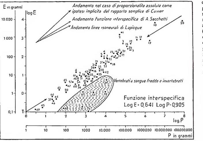
Using an orthogonal graph in which body weight is represented on the abscissa and encephalic weight on the ordinate, one obtains a pattern of values (if logarithms are used) that can be interpolated by an ascending straight line, from the low to the high values, for the mass of mammals and also for various birds (A. Sacchetti). The accompanying graph also shows the straight lines implicit in the procedures previously used by Cuvier and Lapicque, but which have been disproved by the facts as an interspecific relationship. The position of man (with points 1 and 2) is noteworthy, being very high in terms of the predominance of encephalic weight. This position could be assessed by an interspecific coefficient whose measurement gives a relative indication precisely of encephalic weight, thus making comparisons among the various species possible. Other relationships have been demonstrated between the same two weight measurements within the comparative study not only of species, but also of races or other systematic entities, always considering them as populations of individuals. This methodological principle of research belongs to the orientation of modern demogenetic studies, that is, the genetics of populations (v. BIOMETRIA).
As regards the relations between psychology and the massive development of the brain, it must be said that these exist as one among the many factors in the different encephalic evolution of animals and man, and that they also have a different significance according to the structural and morphological differences of the individual cerebral portions. In any case, a certain relationship exists between encephalic weight and the development of intelligence even when the comparison is limited to man. In idiots, for example, the weight of the brain is unusually low, whereas in men of genius it is very often higher than the average for the population to which they belong (Cuvier’s encephalon weighed 1861 g., Byron’s 1805, Schiller’s 1580). This relationship is not, however, at all constant, owing to the many biological factors that interfere with it and many of which remain inadequately understood by our scientific knowledge.
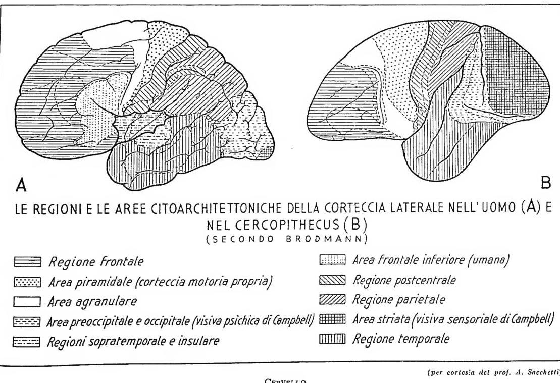
B
(AFTER BRODMANN)
Frontal region
Pyramidal area (proper motor cortex)
Agranular area
Preoccipital and occipital area (Campbell’s psychic visual area)
Supratemporal and insular regions
Inferior frontal area (human)
Postcentral region
Parietal region
Striate area (Campbell’s sensory visual area)
Temporal region
BRAIN
(courtesy of Prof. A. Sacchetti)
Two hemispheres (right and left) are distinguished in the brain, joined at the midline by a horizontal lamina of white matter called the corpus callosum. Each hemisphere has two extremities (one frontal or anterior and one occipital or posterior) and three surfaces: one internal, facing the other hemisphere; one external, facing the cranial wall; and one inferior, facing the base of the skull. The surface of the hemispheres, called the cerebral cortex (cortex), or pallidum, or mantle, is smooth in many animals; hence their name, lissencephalic (R. Owen). In man, on the other hand, as in the higher or gyrencephalic animals, it has numerous protuberances and furrows, the former called convolutions or folds and separated from one another by fissures or sulci.
The presence of these folds indicates considerable development of the grey matter constituting the cortex in relation to the volume of the brain. There are fissures that delimit entire lobes on the individual cerebral surfaces; these are the deepest and most important for phyletic comparisons. Among them are the fissure of Sylvius and that of Rolando. The frontal lobe is situated precisely anterior to the fissure of Rolando and thus has very clearly defined limits. There are also a parietal, an occipital, and a temporal lobe. An enormous body of literature by anatomists and anthropologists deals with the various convolutions of these individual lobes and their phyletic, racial, or physiological significance. The insular lobe, covered by the lips of the fissure of Sylvius, is also of interest in this connection.
The general structure of the cortex is characterized by layers in which fibres and nerve cells of various types alternate. The nerve cells, however, constitute the essential element of the grey matter, which has an average thickness of approximately 2.5 mm. or more. In general, six different superimposed layers are distinguished according to the classification accepted today (Economo, Brodmann, Cajal, etc.); among them, pyramidal, fusiform, and granular cells predominate. The first two categories belong predominantly to projection pathways; the last, which connects larger elements and is provided with shorter neurites, mainly performs associative functions, especially in the sixth layer. This cellular differentiation corresponds to a profound differentiation of nerve fibres that are likewise stratified (Elliot Smith and O. Vogt), as well as to a different appearance among the areas of the individual lobes. The areas that are morphologically distinct and correspond to a different significance in functional localization may be classified as projection areas—
CERVELLO - Distribution of individual species as a function of encephalic weight (log E) and body weight (log P). In a hatched area are reported the values relating to a few cases of cold-blooded vertebrates and invertebrates. The arrows converging toward the centre of the distribution (based for the most part on data from mammals) represent the course of the interpolating function of the same data. Separately, at the upper left of the graph, three arrows are shown according to the respective course implicit in Cuvier’s method, in our interspecific function, and in Lapicque’s system. 1. Homo sapiens L. ♂ 2. Homo sapiens L. ♂ 3. Simia satyrus L. ♂ 4. Hylobates syndactylus Desm. ♂ 5. Hylobates leuciscus Kuhl ♂ 6. Semnopithecus maurus Desm. ♂ 7. Macacus cynomolgus L. ♂ 8. Midas rosalia L. ♂ 9. Nycticebus tardigradus L. ♂ 10. Elephas indicus L. ♂ 11. Equus caballus L. 12. Equus asinus L. ♂ 13. Tapirus americanus L. ♂ 14. Capra hircus L. 15. Oryx beisa Rupp. ♂ 16. Cephalophus Maxwelli H. Sm. 17. Camelopardalis giraffa Schreb. ♂ 18. Tragulus javanicus Pal. ♂ 19. Hippopotamus amphibius L. ♂ 20. Balaenoptera sibbaldi Gray 21. Mustela putorius L. ♂ 22. Lycaon pictus Temm. ♂ 23. Canis familiaris lappon. ♂ 24. Canis familiaris Bernh. ♂ 25. Canis familiaris Bernh. ♂ 26. Canis familiaris Leonb. 27. Viverra civetta Schreb. ♂ 28. Paradoxurus musanga Gray 29. Felis leo L. ♂ 30. Felis concolor L. ♂ 31. Felis domestica Gm. ♂ 32. Lepus cuniculus L. ♂ (forus) 33. Lepus cuniculus L. ♂ (forus) 34. Hydrochoerus capybara Ersi. ♂ 35. Lagostomus trichodactylus Brookes 36. Mus decumanus Pall. ♂ 37. Mus musculus L. ♂ 38. Castor canadensis Kuhl ♂ 39. Sciurus bicolor Sparrm. ♂ 40. Sciurus vulgaris L. ♂ 41. Myrmecophaga rubata L. ♂ 42. Manis javanica Desm. 43. Pteropus edulis Geoffr. ♂ 44. Vespertilio murinus Schreb. ♂ 45. Vespertilio mystacinus Leisl. ♂ 46. Rhinolophus ferrum equinum Schreb. ♂ 47. Tupaia javanica Horsf. ♂ 48. Erinaceus europaeus L. ♂ 49. Sorex vulgaris L. 50. Talpa europaea L. 51. Didelphys marsupialis L. ♂ 52. Thylacinus cynocephalus 53. Macropus 54. Dasyurus maculatus 55. Aquila chrysaetos 56. Buteo vulgaris 57. Falco tinnunculus and F. cenchris 58. Cygnus olor and C. gilvus 59. Anas boschas 60. Anas querquedula 61. Pavo cristatus 62. Phasianus colchicus 63. Minus polyglottus 64. Luscinia rubicola 65. Paleornis docilis 66. Chrysotis amazonicus 67. Alligator mississippiensis 68. Rana catesbiana 69. Cyprinus carpio 70. Cyprinus vulgaris 71. Anguilla vulgaris 72. Octopus vulgaris 73. Homarus vulgaris.
be classified as projection areas (motor areas, with an almost complete absence of true granules), sensory areas (with an enormous richness of granules), and association areas (with lesser development of both types of cells). This conclusion was reached after an infinite series of experimental investigations and clinical and anatomo-pathological observations. It is certainly true, from the physiological point of view, that today the notion of the whole (in this specific case, the c. as a single psychic centre) remains fundamental in biology as in psychology; it is necessary, however, in the light of the facts, to deny that the c., in all its complex structure, participates in every psychic act, even elementary ones. Functional localizations therefore remain a scientifically established fact.
Among the projection centres, for example, there should be noted the tactile sphere, around the fissure of Rolando; the olfactory sphere, on the internal surface; the visual sphere, in the occipital lobe; and the auditory sphere, on the first temporal convolution. Of physiological importance in the frontal lobe (which is especially developed in man) is the inferior convolution of the lateral surface (Broca’s convolution), which governs
(courtesy of Prof. A. Sacchetti)
CERVELLO - Structure of the cerebral cortex (according to Brodmann and Vogt, after Economo); on the left, the six cellular layers (cytoarchitecture); on the right, the corresponding six planes of fibres (myeloarchitecture).
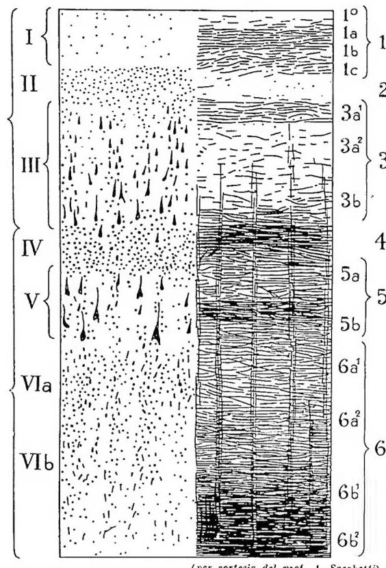
The racial differentiations of the human brain, besides relating particularly to weight and volume, are also macroscopic or microscopic in nature (cytoarchitectonic and myeloarchitectonic). Its general form varies, for example, with the different cranial forms of the races. According to Chiarugi, in brachycephalic human groups the grooves running in an anteroposterior direction would be deeper than in dolichocephalic groups. The indices and measurements of individual encephalic portions would also differ. The fissure of Sylvius would be differently inclined, as also seems to be the case in the fossil men of the Palaeolithic. Other notable differences concern the insular lobe, according to the specific studies of S. Sergi, particularly in comparison with Hylobates syndactylus.
Particular knowledge of the remains of the most ancient men is based on the cubature of the cranial cavity and on the cast of the endocranium. Knowledge concerning the morphology of the folds that left impressions is indirectly based on the latter. These are naturally imprecise and incomplete observations, whose conclusions must be accepted with reservation (for endocranial volume V. PALEANTROPOLOGIA). As regards the grooving, what is striking in the Neanderthals is its general simplicity and the coarse appearance of the convolutions. Particular attention must be paid to the foot of the third frontal convolution, which is certainly present in the Neanderthals, as S. Sergi demonstrated for the Saccopastore skull. In the cast of the Piltdown skull
(lower Palaeolithic), Keith finds a simple and primitive arrangement of the parts, but not so simple and so primitive as to place the Piltdown brain in a class distinct from that of the modern human brain.
The conclusion, with S. Sergi, is as follows: it may be held that, while the brain of the Neanderthals possesses, in the volume and general morphology of the neopallium, the characteristics of living hominids, it differs from them in an ensemble of primitive characters intermediate between those of man and those of the anthropoids. Some differentiating characters even seem to represent a less advanced evolution, «a cui doveva corrispondere una più scarsa attività psichica». This assertion, however, is very uncertain, all the more so because conclusive data are lacking on the encephalic morphology of still more ancient men, such as Pithecanthropus.
Turning to differences in individual variability, among the living, between the individual portions of the brain, the following may be said: eight zones have been identified (A. Sacchetti) with more stable grooving, and without exception they are the most ancient: those that in man have often undergone relative reduction in comparison with the growth of surrounding areas, which are less stable from one individual to another, or with the emergence of new areas and therefore of new grooving and gyrification. This is the case with the olfactory, superior temporal, striate, and limbic areas. All the newly formed areas prove unstable and more variable; fundamentally, they are those that still tend toward a different individual arrangement, those that in man represent a further perfection not only morphological but also physiological and psychological (they are the associative areas), those that give man his so-called personality, not only through the direct cerebral formations but, above all, through the interest they arouse and through the psychological conditions of the higher associative sphere (it would suffice to mention certain frontal areas and the inferior parietal area).
All the data confirm: a) the greater variability of the areas and parts that are more complex from the functional point of view; b) the greater variability of the cellular layers that preside over the characteristic functions of the areas; c) the greater variability of the zones that appear late in individual development and in the course of possible phylogenesis, and that are typically human because of their particular development in man.
Where homologies with the anthropoids and monkeys are more readily established, where we observe the relative reduction of parts in man (for example, by following Tilney’s planimetric coefficients), where there is a relatively lesser complexity and functional specificity, and where formative processes occur early in ontogenetic development, there is generally less variability.
HISTORICAL PRECEDENTS. — In the medical writings which, collected under the name of Hippocrates (468–377 B.C.), form the foundation of ancient medicine, two different concepts of the function of the brain are found. In contradiction to the Homeric belief, which regarded the diaphragm as the seat of spiritual functions, according to Hippocrates “it is through the brain that we become mad, that delirium seizes us, that fear and pain assail us...” ; “We know that pleasure and joy on the one hand, and pains and sorrows on the other, are referred to the brain.” On the other hand, the writings on the cardinal humors regard the brain as the place where mucus, the cold and moist humor (the humor of winter and old age), is prepared. Mucus, descending from the head as “catarrh” or “rheumatism,” is the cause of pneumonia, pleurisy, phthisis, ascites, and diarrhea; while bile is responsible for the heat of fever, mucus causes trembling. Apoplexy and paraplegia are due to mucus, which obstructs the vessels.
The followers of Plato in medicine, the so-called dogmatists, consider the marrow to be the link between body and soul. They see in the brain, perfect in its spherical form, not only the place where semen is prepared but also the seat of reason and the soul, whereas the mortal parts of the soul, such as feelings and desires, are located in the chest and abdomen.
The school of Alexandria, represented by Herophilos (300 B.C.), sees in the brain, and particularly in its ventricles, the seat of the soul. Herophilos is responsible for a highly accurate anatomical description of the brain; and structures such as the calamus scriptorius and torcular still bear his name.
Galen (A.D. 131–200), physician to the Emperor Hadrian, recognized the correlations between psychic and physical factors and professed the doctrine that psychic functions were the expression of the concerted action of the humors. He located in the brain the faculty of drawing conclusions (ἐνυχή λογιστική); emotional life resided in the heart, while desires resided in the liver. Galen, who also devoted himself to the anatomy of the brain, incorporated the ideas of the pneumatic school into his system. According to this school, the pneuma psychicón was formed from the contents of the carotid arteries in the choroid plexuses of the ventricles. Although repeating the Hippocratic ideas concerning the function of mucus, Galen regarded the brain as an insensitive organ, whose movement was coordinated with respiration and whose purpose was to carry the pneuma from the ventricles into the nerves. Galenic ideas dominated medical thought in this field for a millennium and a half.
The Byzantine physician Poseidonios, in the second half of the fourth century, was the first to deny the demonic origin of mental illnesses and to advance the hypothesis that certain cerebral functions were localized. He situated the imagination in the anterior lobe, reason in the central ventricles, and memory in the occipital parts; from this arose the conviction that neuroses and psychoses were due to disorders of these regions.
Arab medicine, represented by Avicenna (980–1037), reflects the ideas of the Greek world and regards psychic alterations as dyscrasias of the brain. The intensity and form of psychoses depend on their localization and severity. Black and yellow bile, as well as putrid mucus, were considered the causes of melancholia, mania, and dementia. As for localization, disorders of the anterior lobe cause hallucinations, those of the central regions cause loss of understanding, while occipital lesions cause loss of memory.
The “School of Salerno,” the principal representative of medieval medical thought, classifies the organs into membra animata, among which the brain predominates; membra spiritualia, dominated by the heart; and membra nutritiva, dominated by the liver. The speculative medicine of the Middle Ages contributed very little to the advancement of scientific knowledge, and only the great anatomists of the Renaissance succeeded in providing a description of the brain superior to that supplied by Galen. The Bolognese Costanzo Varolio (1543–75) described, in 1572, the base of the brain (pons Varolii) with particular accuracy. The professor of Leiden, Franciscus Sylvius (de la Boë) (1614–72), discovered the true structure of the ventricles (aquaeductus Sylvici), and the Swiss Johann Wepfer challenged the formation of animal spirits within them. In 1658, he finally demonstrated that Hippocrates was mistaken and that mucus could not descend from the brain to enter the nasal cavity. The Englishman Thomas Willis (1622–75) gave an accurate description of the cerebral vessels (circuli arteriosus Willisii) and of the corpus striatum in his Cerebri anatome (1664).
In the eighteenth century, knowledge of the brain was advanced by the Frenchman Vicq d’Azyr (1748–94; fasciculus Vicq d’Azyr), and by the German Samuel Thomas Sommering (1755–1830) with his book De basi encephali (substantia nigra Sömmering).
A lively discussion of the functions of the brain, which also engaged the general public, was conducted by Francesco Giuseppe Gall. He acquired indisputable merit by determining the course of the nerve fascicles entering the brain and by describing the decussatio piramidum. In his lectures, begun in 1786, he advanced the theory that twenty-seven mental faculties were recognizably localized on the surface of the brain, such as love of one’s children, the carnivorous instinct, friendship, cunning, wisdom, the metaphysical spirit, poetic talent, etc. This theory, under the name of phrenology, was challenged by official science, and Marie-Jean-Pierre Flourens (1794–1867) deserves credit for laying the foundations of more accurate concepts. By discovering the respiratory center (point vital), he opened the way to research into the vegetative centers of the central nervous system. Among nineteenth-century scientists, Luigi Rolando, with his Saggio sopra la vera struttura del c. dell’uomo e degli animali
(Sassari 1809), Jules-Bernard Luys, and Louis-Pierre Gratiolet, with their comparative anatomy of the brain of humans and apes, opened the way to a deeper understanding—a path which, through histological and physiological discoveries, led to the present picture, marked by the names of Golgi, Martinotti, Baillarger, Gennin, Bechterew, Monakov, Meynert, Edinger, Flechsig, Burdach, Gowers, Deiters, Purkinje, and others.