CRANIUM. – The bony head consists of two distinct portions: the cerebral cranium, which contains the encephalon (v. CERVELLO), and the facial cranium, which contains various organs of the senses and the organs of mastication. The cranium properly so called, or cerebral cranium, is essentially a bony protective case for the most important part of the central nervous axis. Consequently, its study has always been of particular interest from the various anatomical, physiological, anthropological, and clinical points of view. The bones composing it are essentially eight: the frontal, ethmoid, sphenoid, occipital, the two parietals, and the two temporals. Between them, however, there are often intercalated supernumerary ossicles, more or less numerous and developed, called Wormian bones. These bones are classifiable among the flat bones and consist of two plates of compact tissue, one external and one internal, enclosing between them a layer of spongy tissue, highly variable in thickness, called diploë. The internal table (or plate) is in contact with the encephalon and models itself, like wax, over its irregularities. Thus the grooves of the table correspond, as in a negative, to the cerebral convolutions, while the projections (mammillary eminences) correspond to the cerebral sulci. Similarly, on the internal table are found the negative impressions of the arteries and the large venous canals running over the surface of the hemispheres. The study of cerebral morphology through such impressions is, however, subject to many reservations because of the uncertainty of the determinations.
The bones of the facial cranium may be distinguished into two groups, an upper one (the upper maxillary portion) and a lower one (the lower maxillary portion). The lower portion consists solely of the mandible, whereas the upper portion consists of thirteen distinct bones: the upper maxilla, the malar bone (zygomatic), the lacrimal (suguis), the inferior turbinate, the nasal and the palatine bones, all paired and arranged symmetrically with respect to the median plane of the cranium, and the vomer, unpaired. The human cranium is studied descriptively in anatomy, but its interest remains general among anthropologists, who, through the most varied comparisons, seek to establish the differentiating characteristics of the species, race, sex, individual ages, etc. Among the classical names of scholars in the history of anthropology most closely associated with craniological research, the following should be remembered: C. M. Daubenton (1744), G. F. Blumenbach (1775), P. Camper (1791), G. Prichard (1807), and subsequently Geoffroy Saint-Hilaire, F. Cuvier, A. Forville, G. B. Parchappe, G. Serra, P. P. Broca, G. Sergi. But the ranks of researchers could be extended at length, because the morphological and metric study of the bones has always constituted an essential chapter of anthropology (v. ANTROPOLOGIA). This may seem exaggerated, since the human organism is not revealed solely through its bones, but it is understandable for various reasons: in practice, comparative material is more readily constituted by bones, while in the case of fossil humans these are the only remains preserved. Indeed, anthropologists have therefore been confronted with complex problems concerning the origin of human forms and their related transformations, which nevertheless have had to be resolved through skeletal remains. The cranium, naturally, has been the object of the greater part of the research, and in it are in fact revealed the most reliable differentiating characteristics among the most diverse groups, together with traces of the phyletic evolution that took place over time. We shall mention the most important features for racial classification, which also serve as a basis for the study of fossils.
For each of the individual bones composing the cranium, its morphology is defined in relation to group or individual differences; in particular, however, the form of the entire cerebral cranium is studied as viewed according to certain norms, as well as that of the facial cranium in its essential portions.
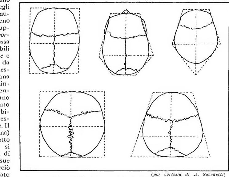
The form of the cerebral cranium viewed from above, vertically at its maximum contour, often serves for a sound racial differentiation, albeit with reservations concerning the first attempts of Giuseppe Sergi, the ingenious initiator of the method. Around 1895 he distinguished
(courtesy of A. Sacchetti)
CRANIO — Diagram of the cranial forms (in the vertical norm) identified by S. Sergi: ellipsoides, ovoides, pentagonoides, sphaeroides, sphenoides.
An example. A Cartesian graph is constructed, ortho-
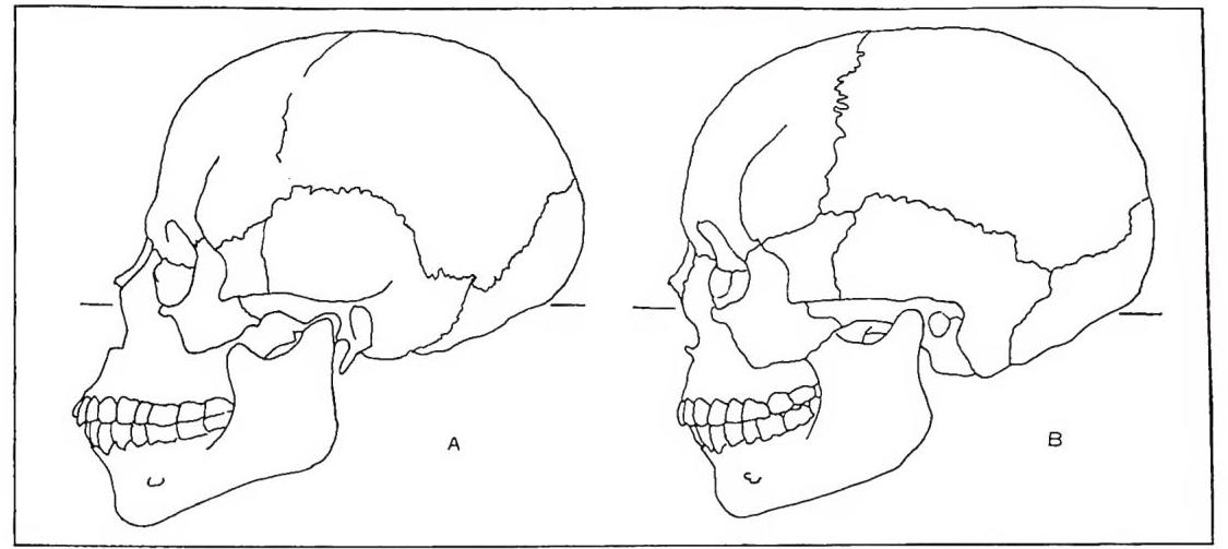
CRANIO - Examples of variation in facial prognathism in two African skulls (diagrams derived from figures by De Quatrefages and Hamy). A) Prognathous skull, a Congolese Negro; B) Orthognathous Hamitic skull. The orientation of the two subjects is based on the orbito-auricular plane of the German Convention.
Among the same metric measurements of the skull, the study of correlations has particular value; it has brought to light racial differences of considerable systematic significance, while clarifying the differing behavior of various cranial regions. The correlation between the length and breadth of the skull has in fact been examined in individual groups of persons (belonging to the most diverse races), and certain positive values have been found, showing that, as length increases, breadth also increases on average from one individual to another. But the same procedure applied to the racial means of several human groups has yielded a zero, and perhaps negative, correlation. This means that the relationship between the two measurements is much more variable among different races than among individuals of the same race. The importance of the result is evident when one considers that there are instead cranial diameters (e.g., those of the facial skeleton) that behave in a completely opposite manner: racial differences in the aforementioned ratios are much smaller than individual differences; in other words, the inter-racial correlation is greater than the intra-racial correlation. This completely opposite behavior among different measurements has been demonstrated only recently by biometric means, contrary to the pessimistic predictions of various authors (v. SACCHETTI, FRANCO).
Among the other craniometric indices most commonly used as indirect measurements of shape, the following should be mentioned: the vertical-longitudinal index (which serves to assess cranial height relatively according to the categories of chamaecephals, orthocephals, and hypsicephals), the frontoparietal index, the total facial index (which serves to assess facial height relatively according to the categories of euryprosopes, mesoprosopes, and leptoprosopes), the orbital index (which serves to assess orbital height relatively according to the categories of chamaeconchs, mesoconchs, and hypsiconchs), and the nasal index (which serves to assess the breadth of the nasal aperture relatively according to the categories of leptorrhines, mesorrhines, and platyrrhines). There are, however, some indices, such as the total facial index, which, although generally used for racial differentiation, have now proved entirely unsuitable for this purpose (with the aforementioned biometric methods) because of their strong individual variability and consequently their extremely low diagnostic value in the field of human systematics. In general, these metric measurements must always be supplemented by a specific morphological analysis of the individual skeletal regions, together with a study of their topographical position in relation to the fundamental planes of orientation of the skull. Anthropologists have devoted particular attention to the latter, for example, in assessing the prognathism of fossil humans. The best results, however, have been obtained on physiological and morphomechanical premises.
Within the same order of research, mention should be made of those concerned with measuring cranial curves and angles (particularly at the base of the skull, on the sphenoid, and in the face), as well as the capacity of the cranial vault, on which observations of great interest have been made because of the corresponding inferences concerning brain mass, especially in the case of fossil humans (v. CERVELLO; PALEANTROPOLOGIA). The most important facial angle is the one that permits the measurement of prognathism and has yielded good results for racial differentiation. It should be noted, however, that prognathism is now also determined analytically through the spatial morphomechanical study of the individual bony elements. Particular attention should be paid to this last point. All modern craniology in fact aims not only to establish the systematic value of characteristics in differentiation, but also to explain physiologically and mechanically the various architectural and structural adaptations of the skeleton. The function of mastication is evidently connected with the alveolar arch and with the arrangement of the individual bony elements, just as the architecture of the cranial vault depends on the spatial position of the temporal bone, which contains the organs of orientation. It has in fact been
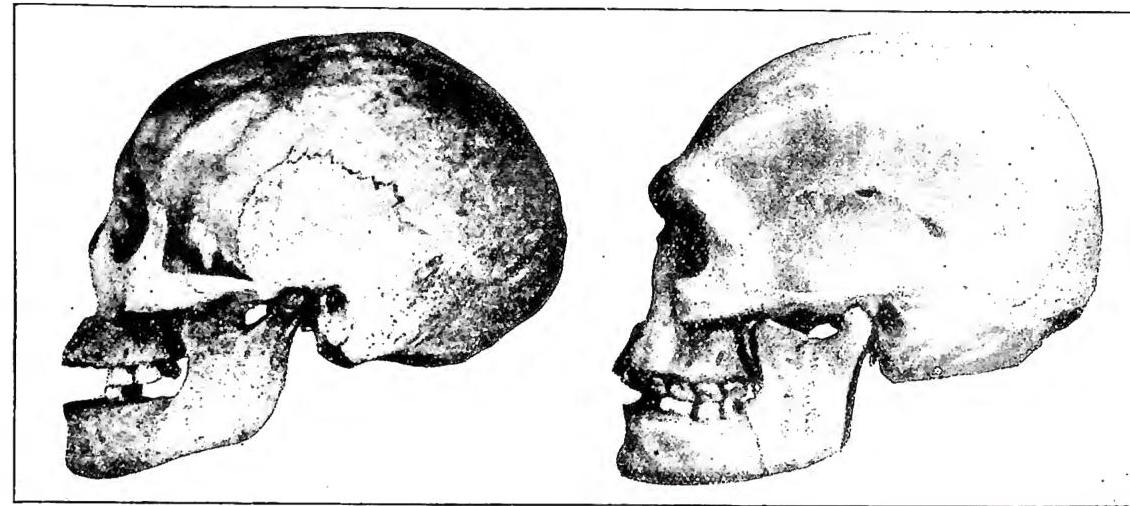
This analysis of the position and form of the individual bones in relation to the cranial solid as a whole and to its physiological planes of orientation is pursued by means of specific techniques and specially constructed instruments (v. ANTROPOLOGIA). The accompanying figures provide an example of the study of the isolated maxilla in relation to the fundamental planes, while the corresponding geometric diagrams, superimposed upon one another, clearly show the differences sought, allowing a more rational interpretation. It should be noted in this regard that the reference points of each bone are those of greatest architectural importance in craniogenesis.
The accompanying figures also show notable examples of racial differences in various other characteristics of the human skull.
There are also morphological and structural differences dependent on sex, which make it possible to distinguish the male skull from the female even at the same age. The female skull is generally said to retain a morphological complex of greater infantilism, which makes it more delicate and thinner than the male skull. It is in fact also smaller and lighter; the bony crests for muscular insertions are less pronounced, the mastoid processes smaller, and the forehead more arched, with a bambée form recalling that of children. The frontal and parietal eminences are more developed, the superior margins of the orbits more sharply defined, almost cutting, and the overall volume of the cranial vault is smaller, etc. Naturally, all these typical characteristics are useful in ordinary cases, but the anthropologist may encounter exceptions in which diagnosis remains highly uncertain, if not impossible. There are cases, in fact, in which female skulls display characteristics of robustness entirely similar to those of males, to the point that they cannot be distinguished, while there are cases of male skulls with delicate features that are completely confused with the corresponding characteristics of the other sex. The norm, however, is that indicated above.
The development of the skull through the ages of life has likewise been studied, so that examination of various characteristics enables the anthropologist to make a reasonably accurate diagnosis of age. Particularly noteworthy are the transformations of the cranial vault in children resulting from the ossification processes that occur after birth, within the so-called membranous skull, which is composed of fibrous tissue in the vault, whereas the cranial base is constituted at an early stage during the embryonic period by cartilage (basilar plate of Kölliker). In the child’s membranous skull (the fontanelle period), numerous ossification centers are observed, which gradually form the bones of the vault. Consequently, the angles of the cranial bones, being farthest from the original ossification centers, are the last to be filled by osseous substance. For this reason, the corresponding regions received the name fontanelles. They are thus of interest both to the obstetrician in prenatal diagnosis and to the anthropologist, who observes the different developmental patterns among the most diverse races. With the disappearance of the fontanelles through complete ossification of the vault, the margins of the individual bones assume a serrated form, permitting a kind of reciprocal inlaid interpenetration of the adjoining elements. The complexity of these serrations at the so-called sutures of the skull varies from region to region, but generally increases with age, although at different rates among the various racial groups. All these elements are valuable for diagnosing the age of individuals. The specialized anthropologist can nevertheless distinguish the not uncommon cases of anomalous obliteration of the sutures as the final stage of synostosis. The membrane remaining at the sutures is gradually invaded by osseous tissue (as previously occurred with the fontanelles), so that the various parts of the skull, albeit at different times, eventually fuse completely with one another. Here too, naturally, one must distinguish a normal physiological process, which leads to the condition of the classic skull of an old person, consisting of a single bony piece, from a pathological process that may even produce major abnormalities of cranial form (microcephaly, scaphocephaly, plagiocephaly, etc.). It has also been debated whether a different progression in the obliteration of the cranial sutures might explain the various cephalic forms according, for example, to the diagrams of G. Sergi. Perhaps, however, this is only one of the dynamic factors in the craniogenesis of forms—a factor that may nevertheless enlighten us when we seek to interpret their nature and immediate biological causes, at least in individual ethnic groups.
In conclusion, the human skull differs according to age, sex, and race, in ways that are still being studied through modern methods of statistical and anthropological analysis. Very often, the ultimate racial differences have not been explained in their nature, and their temporal evolution is unknown. The so-called connecting links between the different forms are lacking. The problems, however, are being examined in increasing depth, and research is tending ever more to clarify the modes of behavior of individual characteristics through the generations, as well as in cases of miscegenation or hybridism and under the most varied environmental influences. The problem of racial mixtures is fundamental and often explains the genesis of new forms. Particular mixtures of different types sometimes serve as indirect evidence for observing the architectural adaptation of specific bony structures. The example in the figure, in which two maxillary diagrams from Arbore individuals are superimposed, was used precisely in the study to explain the facial metamorphism of those populations living on Lake Stefania, at the boundary between the inhabited world of the Negro peoples and that of the Hamitic peoples, among whom, conversely, European facial forms predominate. Finally, the problems of the plasticity of characteristics as a function of the environment have also been studied in the case of the skull, especially with regard to its general form according to the concept of the cephalic index. The investigations of F. Boas on immigrants in America are well known, as are the data of Snigrew and Talko-Hryncewicz for Lithuania, of Fishberg for New York, of Elkind for Warsaw, of Zakrewsky, Majer, and Kopernicki for Galicia, of Hirsch for Massachusetts, of Guthe for Boston, as well as the investigations of Frets, Bryn, Schreiner, Geipel, Abel, and Pessler, the more recent studies of Dornfeldt, and those of Sacchetti. The conclusions that appear most probable, according to the latter, suggest a norm of reaction of individual characteristics, such as the shape of the head, in the strictly genetic sense of the expression and in cases of transformations established following a change of environment (through the emigration of entire groups, etc.). In particular, the cephalic index, in comparison with all the other anthropometric characteristics of the bony head, has shown great and significant inter-racial variability; this result might appear to conflict with the aforementioned knowledge of the possible variations of the characteristic following environmental stimuli. But a careful analysis of the phenomena shows that even those small variations in the shape of the head, which so greatly concerned anthropologists at the beginning of the century (causing them to divide into two opposing camps over whether to admit or exclude environmental influences), should not be considered contrary to the very aptitude itself
of the character toward the differentiation of groups, still less with their actual differing morphological and demogenetic constitution, if one considers the strictly genetic and physiological aspect connected with II. This involves a principle of reactivity linked to the individual genetic endowment and manifested through the phenotype. Only in this way can it be explained how, when faced with the same social and geographical environment (e.g., in America, U.S.A.), racially different populations (brachycephalic Polish Jews and dolichocephalic Mediterranean Sicilians) reacted in opposite ways to one another, irrespective of the individual forms of their intermixture with the local populations (Boas). Something similar occurred with certain groups of emigrants in Italy (Sacchetti). Reactivity to the environment and heredity thus do not appear as two contrasting concepts, at least with regard to certain characteristics of the c. in anthropology. Similar examples, however, are already cited in various fields of application of genetics, particularly demogenetics. Thus, to the static and exclusively morphological conception of the study of the human c. there has today been added a new method of investigation in the field of demogenetic determination, always in relation to the reciprocal mechanical adaptation of forms; in this way we approach ever more closely and effectively the explanation of the real, complex phenotypic expression of human groups as they are, within their natural environment and in their potential reactivity to external factors.