GAMBETTA, LÉON-MICHEL. — French politician, born on 2 April 1838 in Cahors, died on 31 December 1882 at Ville-d'Avray (Seine-et-Oise). A descendant of a Genoese family that had settled in France at the beginning of the century, G. devoted himself ■ in Paris to law and revealed himself as an orator before the wider public during the trial of the Réveil.
The editorial staff of the Réveil had been referred to the judicial authorities for having promoted a subscription for a monument to a deputy who had fallen on the barricades in 1851 while opposing the coup d’état of the future Napoleon III. Against the Napoleonic Empire, G. was a determined representative of the republican opposition. After the fall of the Second Empire, he ardently contributed to the consolidation of the Third Republic, promoting an anticlerical policy (« Le cléricalisme, voilà l'ennemi! »), in order to oppose attempts at monarchical restoration and to avoid international complications that might arise from a position clearly favorable to the Holy See in the Roman question. President of the Council of Ministers (14 November 1881), he presented Parliament with various bills that, in his view, were to regulate relations between Church and State, but his grand ministère lasted barely 66 days, and his bills, tending toward the evident dependence of the Church on the State, did not have time to be approved.
Silvio Furlani
GAMBIA, « SUI IURIS » MISSION. — It is situated in the eponymous colony of British West Africa at the mouth of the G. River; it borders the apostolic vicariate of Dakar and the apostolic prefecture of Ziguinchor. It shares with the missions of Senegambia and Senegal (today the apostolic vicariate of Dakar and the apostolic prefecture of S. Louis of Senegal) the history of its evangelization, having been detached only on 6 May 1931 from what was then the apostolic prefecture of Senegal.
It has a tropical and malarial climate, an area of 10,400 sq. km, and a population of approximately 300,000 of the Sudanese Negro race. The Ordinary’s see is Bathurst. It has (in 1948) 3,355 indigenous Catholics, 100 foreigners, and 100 of mixed descent, 718 catechumens, 16 Eastern-rite dissidents, 3,500 Protestants, 2 Jews, 101,274 Muslims, 21 catechists, 17 male teachers and 14 female teachers, 20 male baptizers and 4 female baptizers, 4 principal and 21 secondary mission stations, 2 churches and 4 chapels, 6 elementary schools with 683 pupils, 2 secondary schools with 215 pupils, one vocational school with 15 female pupils, 2 schools for catechists with 24 pupils, and 16 prayer schools with 600 pupils. Associations of Catholic Action are present.
(properly Enc. Catt.)
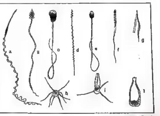
GAMETI ■ GAMETOGENESIS — Fig. 2. Spermatozoa: a) Triton; b) Nereia (annelid); c) Cevia; d) Phyllopneust (bird); e) Man; f) Bat; g) Castrada (turbellarian); h) Pinnoterea (crustacean); i) Lobster; l) Ascaris (worm).
of the germ line), and sometimes already at the stage of two cells (blastomeres) the one that will give rise to the gametes can be identified; but more often, as in mammals and humans, this difference cannot be detected at an early stage.
In the latter case there are male gametes, or spermatozoa or sperms, and female gametes, or eggs. If the difference consists only in size, they are designated as macrogametes and microgametes. Whereas in cases of physiological anisogamy all gametes are motile, as in isogametes, because they possess flagella that permit them to encounter one another for the formation, through the copulative act, of the zygote (v. FECONDAZIONE), in the case of morphological anisogamy there is a motile element, the sperm, and a sta-
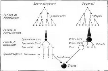
GAMETI E GAMETOGENESI - Fig. 3. Diagram of the maturation of germ cells or gametes (diploid cells in black; haploid cells in white).
whereas in the case of morphological anisogamy there is a motile element, the spermatozoon, and an immobile element, the egg. This difference is related to the egg’s need to accumulate a certain quantity of reserve material, the yolk, which will serve the embryo’s energy requirements during the early stages of development. The accumulation of yolk is more or less abundant in different animals, depending on their particular ecology and that of their juvenile forms (according to whether the eggs are laid in water or on dry land, according to the duration of embryonic life, according to the presence or absence of a larval stage, etc.); consequently, the size of eggs varies greatly among the different animal groups.
According to the quantity and distribution of yolk, eggs are called oligolecithal or alecithal if, like the human egg, they are poor in yolk and therefore very small; they are called telolecithal if yolk is abundant. These are further divided into holoblastic telolecithal eggs, if the entire egg undergoes segmentation (as in amphibians), and meroblastic telolecithal eggs, if segmentation is partial while the yolk mass remains unsegmented at one pole of the cell (as in selachians, reptiles, and birds); a particular case of this type of egg is the centrolecithal egg of arthropods, in which, after segmentation, the yolk accumulates at the centre of the egg (fig. 1).
The egg may possess only its own cell membrane (alecithal eggs), or a membrane and shells added to it during passage through the maternal oviducts (cleidoic eggs).
The spermatozoon, or spermium, is instead characterized by its motility. It is a very small cell consisting of a head, containing the nucleus, a cytoplasmic intermediate piece, and a very long tail that serves as a propulsive organ. The head also contains a cytoplasmic organelle, the acrosome, often characteristically formed into a sharp structure called the perforatorium; sometimes, in addition to the flagellated tail, there is also a more or less extensive undulating membrane.
The spermatozoa of the various animal species have typical structural characteristics by which they can be distinguished (fig. 2). In addition to these typical flagellated spermatozoa, there are others without a flagellum, whose movement occurs through pseudopodia and outgrowths; characteristic are those of an amoeboid form in certain worms and those furnished with various appendages in many crustaceans (fig. 2). There are also cases of immobile spermatozoa that are taken up by eggs through pseudopodia.
The process by which gametes develop into spermatozoa and eggs capable of fertilization is called gametogenesis, and is therefore divided into spermatogenesis and oogenesis (fig. 3).
The primordial germ cells, not yet differentiable into male and female cells, are called protogonia; they are termed spermatogonia and oogonia when the subsequent production of spermatozoa or eggs can be identified. After a period of multiplication of the spermatogonia and oogonia, of more or less long duration and generally much shorter for the oogonia, the cells enter a period of growth and are called spermatocytes and oocytes. At the end of growth, very brief for spermatocytes and longer for oocytes because vitellogenesis occurs during this period, the stage of the first-order spermatocyte and first-order oocyte is reached. From this stage begins the maturation period, which consists in the reduction of the nucleus from the diploid condition, created at fertilization in the zygote, to the haploid condition characteristic of mature germ cells (v. CROMOSOMA). This process takes place through two successive cell divisions of the spermatocyte or oocyte, so that in the end each cell gives rise to four cells. In the nucleus, however, there is only one longitudinal division of the chromosomes (v. RIPRODUZIONE), and therefore a distribution among the four cells of the number thus doubled, so that each consequently possesses half the original number. Thus, from the diploid condition 2n, the haploid condition n is obtained. The first division of the first-order spermatocyte produces two second-order spermatocytes, and the second division produces four spermatids. The first division of the first-order oocyte produces a second-order oocyte and a polar body, or polocyte; the second-order oocyte produces a mature egg and a second polocyte. The first polocyte sometimes divides into two polocytes (fig. 3). The maturation phase of the germ cells is called meiosis.
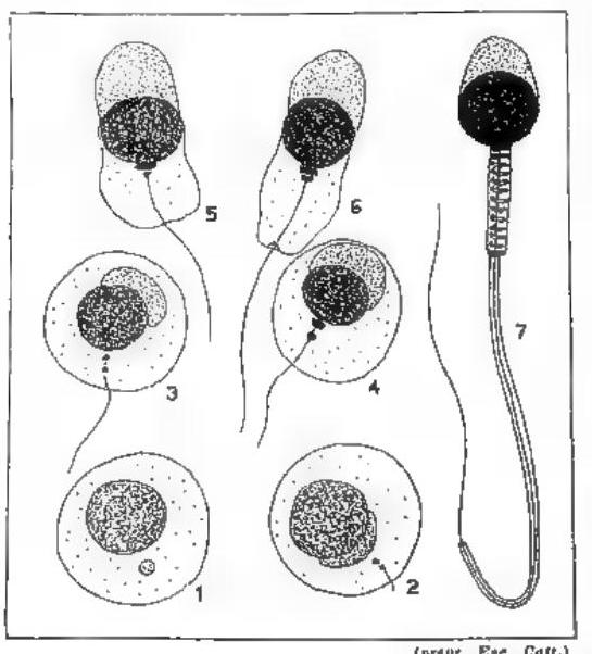
While the mature egg is ready for fertilization, the spermatids, which have the appearance of typical cells, must undergo functional maturation; that is, through a transformation called spermiogenesis, they must become flagellated spermatozoa capable of movement (fig. 4).
The mechanism of gamete maturation shows that four spermatozoa mature for every egg. The percentage difference between the gametes of the two sexes is further increased in favour of the spermatozoa by the activity of the spermatogonia, which continues for a much longer time. In the human species, the activity of the oogonia ceases during the fetal period, before birth, whereas the male’s spermatogonia continue to multiply until advanced age.
This enormous number of mobile male cells is related to the need to ensure the meeting of the spermatozoon with the egg for the preservation of the species.
In the particular case of humans, the numerical ratio between eggs and spermatozoa is 1 to 850,000,000.